| |
Bacteria under Microscope
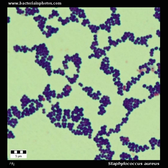
Staphylococcus aureus
|
|
|
| |
Gram-stain: |
Gram-positive |
| |
Microscopic appearance: |
Cocci in grape-like clusters |
| |
Clinical significance: |
- Frequently found as part of the normal skin flora on the skin and nasal passages
- It is estimated that 20% of the human population are long-term carriers of S. aureus
- Skin infections (pimples, impetigo, boils (furuncles), cellulitis folliculitis, carbuncles, scalded skin syndrome, abscesses)
- Postsurgical wound infections
- Pneumonia
- Meningitis
- Osteomyelitis
- Endocarditis
- Toxic shock syndrome (TSS)
- Bacteremia
- One of the five most common causes of nosocomial infections
- Methicillin resistant Staphylococcus aureus (MRSA) infections
|
| |
|
Text: Wikipedia |
| |
Colony morphology: |
|
|
| |
|
|
|
|
|
A |
B |
C |
|
| Staphylococcus aureus identification |
|
A |
Beta-hemolytic colonies of Staphylococcus aureus on sheep blood agar. Cultivation 24 hours, aerobic atmosphere, 37°C. |
|
|
B |
Yellow colored colonies of Staphylococcus aureus on Tryptic Soy Agar. Carotenoid pigment staphyloxanthin is responsible for the characteristic golden colour of S. aureus colonies. This pigment acts as a virulence factor.
Cultivation 24 hours in an aerobic atmosphere, 37°C. |
|
|
C |
Colonies of Staphylococcus aureus seen with transmitted light. Cultivated on Columbia agar with 5% defibrinated sheep blood, 24 hours in an aerobic atmosphere, 37°C.
Colonies are surroundend by a wide zone of beta-hemolysis. |
|
|
|
| |
www.bacteriainphotos.com |
|
|